Learn about coral reefs and giant corals
Giant corals: How they live, their habitat, their health and competing organisms
This section will help you gain some knowledge on the reefs and on corals highlighting those features that might be useful during your mission to locate Giant Corals.
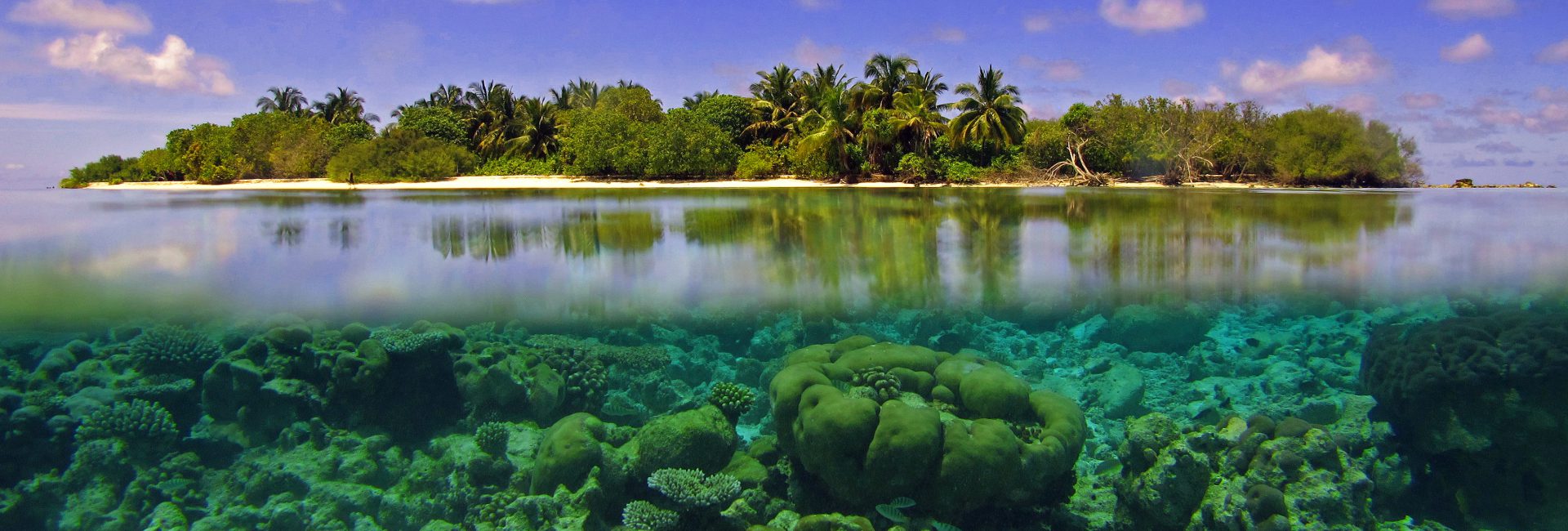
Coral reefs and corals
Coral reefs cover less than 1% of the ocean surface, yet they support 25% of the known marine life. They are living structures made of hard corals, animals belonging to a group of invertebrates called Cnidaria.
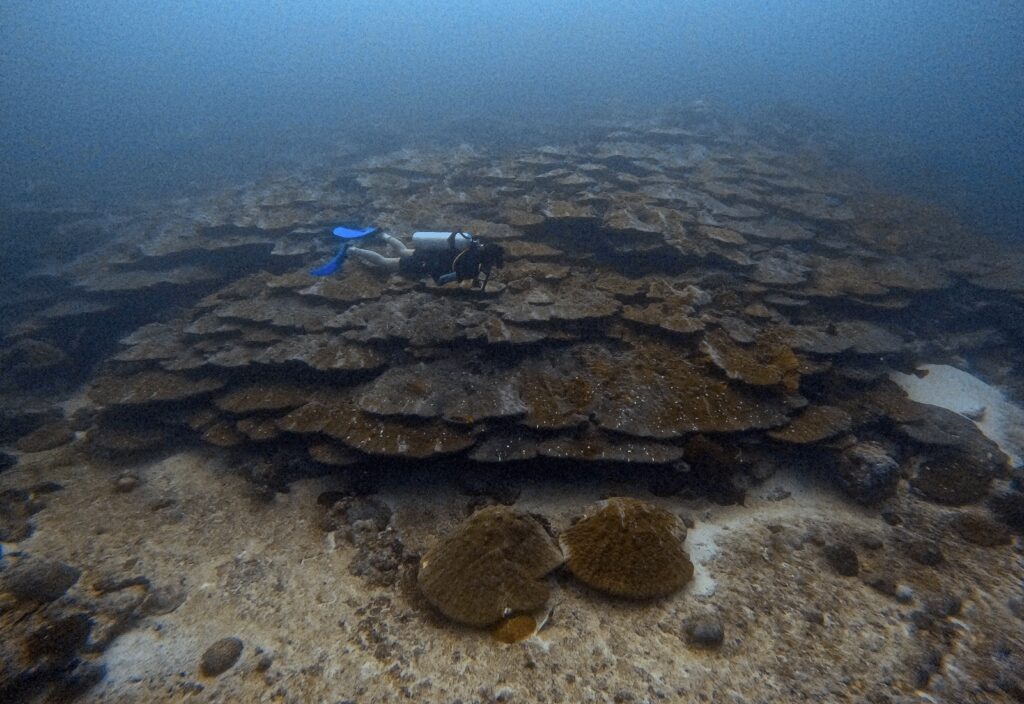
Aside from a few genera, most of them form colonies made of thousands of polyps growing a hard calcium carbonate skeleton and creating coral reefs in the tropical areas of the whole world. A colony originates either via sexual reproduction or from the fragmentation of a larger coral. Colonies then grow in size as each polyp divides itself into daughter polyps, thus potentially growing indefinitely. In American Samoa several very large coral colonies have been reported. The most famous, “Big Momma”, is a Porites sp. measuring over 6 meters in height, 41 meters in circumference, and likely over 500 years old (Brown et al., 2009). The largest Porites sp. in the area was reported by Coward et al. (2020) measuring 69 m in circumference, 22.4 m in diameter and 8m in height.
In November 2024, an incredibly large Pavona sp. has been reported by National Geographic Pristine Seas, from the Solomon Islands, measuring 34 meters wide, 32 meters long and 5.5 meters high. Possibly one of the largest, if not not the largest to date.


Growth forms and growth
Giant corals, and corals in general, can grow in different forms related to their most common reproductive method and lifespan. Terms to describe them are very evocative, allowing for easy grouping:
- Massive corals: massive refers to their dense shape and hemispherical to dome shaped. They are slow-growing species, sometimes growing between 4 and 10 mm/year (Dullo, 2005), and although they can reach large sizes, this is not always the case. They include corals like Porites spp. and Diploastrea heliopora that grows even slower than Porites (Canesi et al. 2024)
- Columnar corals: they form columnar cylinders which are not branching out, such as Pavona clavus or Psammocora sp. For example Pavona clavus colonies from Costa Rica are reported to grow between 6.1 ± 2.9 mm /year (Gateño et al,, 2003) or 9.6 ± 1.77 mm/ year (Guzmán and Cortés, 1989).
- Foliose corals are thin corals flattened horizontally and can form whirl-like structures made of several unifacial plates. For example, Turbinaria spp. might grow around than 1.3cm a year (Browne, 2012) .
- Table corals: they have a column that holds them up on the ground above which they grow horizontally with table-like surfaces, for example some Acroporids. In some cases, these fast-growing, yet vulnerable corals can reach remarkable sizes.
- Encrusting corals: they grow following the substrate they adhere to, forming a thin crust of skeleton over it.
- Free-living corals are made of single polyps or colonial organisms not attached to the substrate.
- Branching corals: they form thin tree-like colonies growing upwards and generally showing a fast growth rate, e.g., up to 150mm/year in the Indo-Pacific (Dullo, 2005). They are currently not considered within giants reports.


Giant corals
Despite ever increasing threats for coral reefs, and despite the average life-span of most scleractinian corals is limited (Devlin-Durante et al., 2016), some coral colonies demonstrated exceptional longevity and growth capacity, reaching impressive ages and sizes (Bythell et al., 2018). The longevity of giant corals might be due to species and individual (genetic) traits or to local conditions. In fact, certain colonies might exhibit genetic resistance (Drury, 2020), conferring them exceptional tolerance to anthropogenic stresses (Sweet and Brown 2016), or to climate change impacts (Mellin et al., 2024), as well as an increased resilience to a changing environment (Coward et al. 2020) permitting them to recover after impacts. Additionally (or alternatively), their existence might be owed to local and regional environmental conditions. That is, giant colonies might have benefitted from inhabiting remote locations, or refugia habitat which, so far, have been spared by destructive global change impacts (Hoegh-Guldberg and Bruno, 2010)
Habitat
Scleractinian corals can be found in tropical areas of the world where temperatures range between 20 to 32°C (depending on different regions). They also need light to grow since they host photosynthetic symbionts in their tissue (algae called zooxanthellae). Through their limestone skeletons, they can build giant bioconstructions even forming islands, atolls, and archipelagos (such as the Maldives). Corals create different habitats that host thousands of marine species. Generally, certain coral growth forms are generally more common than others in each area, depending also on hydrodynamics, light availability, and sedimentation. Within coral reefs, different areas with defined boundaries can be found:
- Lagoon: areas of calm and relatively shallow waters inside the main reef not exposed to oceanic waves. Sometimes devoid of corals or presenting several patch reefs
- Reef flat: a large and shallow, mostly barren area strewn with detritus with an elevation varying very little
- Reef crest: a shallow and highly exposed zone characterised by the presence of coralline algae and mostly encrusting corals. In more sheltered environments more growth forms can be present
- Fore reef slope: this zone extends from the reef crest to the shelf break. It is generally exposed to waves being the outermost zone of the reefs. The mesophotic zone of the reef begins from 30-50m to 300m.
Health and competing organisms
Coral reefs are under threat, and with them, we might lose some of the oldest and most resistant colonies, giant corals. IUCN Red List of Endangered Species has highlighted how reef-forming scleractinian corals are the category (amongst birds, mammals, amphibians and cycads) which is moving faster towards a trajectory of extinction (IUCN 2020) with over 40% of coral species being threatened (IUCN 2024). They are projected with high confidence to decline by 70-90% with a 1.5°C global warming, and there is a high likelihood of species extinction and biodiversity loss (IPCC 2023). In particular, coral reefs are extremely vulnerable to climate change with risks deriving from higher water temperatures, increased severity of storms, ocean acidification, and possible phase shifts (from coral-dominated reefs to algal-dominated reefs) reducing the potential for reef recovery.
In the Maldives, where our project began, global threats coupled with local threats such as land reclamation (Fallati et al. 2017), coral diseases (Montano et al. 2015) and corallivores outbreaks (Bruckner & Coward, 2018; Saponari et al. 2018, Montalbetti et al. 2019). In recent years, Maldivian coral reefs have seen major declines in health and coral cover. Before the 1998 mass bleaching event, reefs averaged 40% coral cover and dropped to less than 2% in 1998 (Pisapia et al., 2016) to then recover to almost 38% in 2009 (Pisapia et al., 2016) and presented a coral community similar to pre-1998 by 2014 (Morri et al., 2015). Following 2016, bleaching event reports indicate communities with 6-11% coral cover (Perry & Morgan, 2017; Pisapia et al., 2019) and changes in the dominant coral genera. In certain locations, massive corals have become the dominant growth forms after bleaching events due to their resistance. Nonetheless, they are subject to possible bleaching, diseases and predation by competing organisms.
Bleached corals appear white in colour, with tissue present on the skeleton and transparent polyps. Bleaching results from disrupting the symbiosis between coral polyps and the photosynthetic microscopic algae (zooxanthellae) found within their tissue. These are responsible not only for corals coloration but also for providing them with most of the energy necessary for their growth. In high temperature conditions, the symbiosis is broken and coral might either die of heat or starve to death unless the original conditions are restored.


Predation generally leaves scars on the colony and most of the time predators are found nearby. Not all predators cause permanent damage to corals (especially giants) and amongst these, we can mention a few: the Pin-cushion Seastar (Culcita sp.), worm snails in the family Vermetidae (Dendropoma spp.), different species of sponges, and certain species of fish biting off pieces of the colonies e.g., parrotfish (Scaridae), damselfish (Pomacentridae), Triggerfish (Balistidae), Butterflyfish (Chaetodontidae). Others like the Crown of Thorns Seastar (Acanthaster planci), or the sea snail Drupella sp. can instead have large-scale impacts. Outbreaks of them have caused in the Maldives (e.g., Bruckner & Coward, 2018) and other parts of the world (e.g., Comming, 2009) large-scale mortality events targeting preferably branching corals, but also moving towards massive corals following the lack of branching species post-bleaching events.
In terms of competing organisms, accounts of the sponge Terpios hoshinota have been reported from the Maldives (Allison, 2024) and the organism is known for overgrowing corals and undergoing outbreaks.


Predators have been reported to have several effects on corals: they can cause complete or partial mortality of corals (Rotjan & Lewis, 2008), be the vector of a disease or leave lesions that may cause infections (Nicolet, 2018). The first report of coral diseases in the Maldives dates back to 2012 (Montano et al., 2012) and since then five diseases have been found in the region (Montano et al, 2016). Given the slow-growth of certain coral morphologies and the mass mortality that diseases are causing (Walton et al. 2018), key to conservation is the early detection of any disease.
The most common coral diseases are:
- Stony Coral Tissue Loss Disease (SCTLD) , is a highly destructive coral disease that has been significantly impacting coral reefs, particularly in the Caribbean since its emergence around 2014. The primary symptom is a rapid and widespread tissue loss that leaves behind a white skeleton. It affects mostly massive species with mid-to-large sizes, dense skeletons, low growth rates, and broadcasting sexual reproduction (Alvarez-Filip et al., 2022)
- Skeletal Eroding Band (SEB), recognised by black specks clustered within the corallites of a bare skeleton
- Black Band Disease (BBD), identified by a dark band between live tissue and exposed white skeleton
- Brown Band Disease (BrB), showing a brown band found between a narrow white stripe following the live tissue, and the white skeleton
- White Syndrome (WS), is a common term used to describe acute tissue loss in Indo-Pacific corals which are not showing other disease signs and have a non-established causation. In the Caribbeam similar signs of tissue loss are also present and several diseases described with various names (e.g., White Plague, White band…). Beeden et al. (2008) describe it as “Diffuse patterns of tissue loss that expose bands or patches of bare white skeleton abutting live tissue.” There might be a “visible colour gradient from bare white skeleton to brown as fouling community develops – indicates progressive tissue loss;” and “the margins of lesions may be linear, irregular or annular (ring-like)”


Published scientific articles on Map the Giants
Montano S, Siena FM, Strona G (2024) Finding millennia-old ‘monumental’ corals could unlock secrets of climate resilience. Nature 629:286. https://doi.org/10.1038/d41586-024-01342-8
Siena, F. M., Galli, P., Fallati, L., Dehnert, I., Gobbato, J., Afzal, M. S., Strona, G. & Montano, S. (2025). Mapping monumental corals through citizen science. Coral Reefs, 1-11. https://doi.org/10.1007/s00338-025-02688-9
Read it here: https://rdcu.be/ergh2
References
Allison, W. R. (2024). The ecology of Terpios hoshinota in the Maldives based on three decades of observation. Galaxea, Journal of Coral Reef Studies, 26(1), 27-42. https://doi.org/10.3755/galaxea.G26N-6
Alvarez-Filip, L., González-Barrios, F. J., Pérez-Cervantes, E., Molina-Hernández, A., & Estrada-Saldívar, N. (2022). Stony coral tissue loss disease decimated Caribbean coral populations and reshaped reef functionality. Communications Biology, 5(1), 440. https://doi.org/10.1038/s42003-022-03398-6
Beeden, R., Willis, B. L., Raymundo, L. J., Page, C. A., & Weil, E. (2008). Underwater Cards for Assessing Coral Health on Indo-Pacific Reefs. CRTR Program Project Executing Agency. Center for Marine Studies. The University of Queensland. Australia.
Brown, D. P., Basch, L., Barshis, D., Forsman, Z., Fenner, D., & Goldberg, J. (2009). American Samoa’s island of giants: Massive Porites colonies at Ta’u Island. Coral Reefs, 28, 735-735. https://doi.org/10.1007/s00338-009-0494-8
Browne, N. K. (2012). Spatial and temporal variations in coral growth on an inshore turbid reef subjected to multiple disturbances. Marine Environmental Research, 77, 71–83. https://doi.org/10.1016/j.marenvres.2012.02.005
Bruckner, A. W., Coward, G., Bimson, K., & Rattanawongwan, T. (2017). Predation by feeding aggregations of Drupella spp. inhibits the recovery of reefs damaged by a mass bleaching event. Coral Reefs, 36(4), 1181-1187. https://doi.org/10.1007/s00338-017-1612-6
Bythell JC, Brown BE, Kirkwood TB (2018) Do reef corals age? Biol Rev 93(2):1192–1202. https://doi.org/10.1111/brv.12391
Canesi, M., Douville, E., Montagna, P., Bordier, L., Caquineau, S., Pons-Branchu, E., Iwankow, G., Stolarski, J., Allemand, D., Planes, S., Moulin, C., Lombard, F., Bourdin, G., Troublé, R., Agostini, S., Banaigs, B., Boissin, E., Boss, E., Bowler, C., … Reynaud, S. (2024). Sea surface temperature reconstruction in the Pacific Ocean using multi-elemental proxy in Porites and Diploastrea corals: Application to Palau Archipelago. Chemical Geology, 645, 121884. https://doi.org/10.1016/j.chemgeo.2023.121884
Comming, R. L. (2009). Population outbreaks and large aggregations of Drupella on the Great Barrier Reef. Great Barrier Reef Marine Park Authority.
Coward, G., Lawrence, A., Ripley, N., Brown, V., Sudek, M., Brown, E., … & Vargas-Ángel, B. (2020). A new record for a massive Porites colony at Ta’u Island, American Samoa. Scientific Reports, 10(1), 21359. https://doi.org/10.1038/s41598-020-78484-9
Devlin-Durante MK, Miller MW, Precht WF, Baums IB, Caribbean Acropora Research Group (2016) How old are you? Genet age estimates in a clonal animal. Mol Ecol 25:5628–5646. https://doi.org/10.1111/mec.13865
Drury C (2020) Resilience in reef-building corals: The ecological and evolutionary importance of the host response to thermal stress. Mol Ecol 29(3):448–465. https://doi.org/10.1111/mec.15337
Dullo, W. C. (2005). Coral growth and reef growth: A brief review. Facies, 51(1), 33-48. https://doi.org/10.1007/s10347-005-0060-y
Fallati, L., Savini, A., Sterlacchini, S., & Galli, P. (2017). Land use and land cover (LULC) of the Republic of the Maldives: First national map and LULC change analysis using remote-sensing data. Environmental Monitoring and Assessment, 189(8), 417. https://doi.org/10.1007/s10661-017-6089-2
Gateño, D., Leon, A., Barki, Y., Cortes, J., & Rinkevich, B. (2003). Skeletal tumor formations in the massive coral Pavona clavus. Marine Ecology Progress Series, 258, 97-108. https://doi.org/10.3354/meps258097
Guzman, H. M., & Cortes, J. (1989). Growth rates of eight species of scleractinian corals in the eastern Pacific (Costa Rica). Bulletin of Marine Science, 44(3), 1186-1194.
Hoegh-Guldberg O, Bruno JF (2010) The impact of climate change on the world’s marine ecosystems. Science 328:1523–1528. https://doi.org/10.1126/science.1189930
Intergovernmental Panel on Climate Change (IPCC). (2023). Climate change 2023: Synthesis report. Contribution of Working Groups I, II, and III to the Sixth Assessment Report of the Intergovernmental Panel on Climate Change (H. Lee & J. Romero, Eds.).
International Union for Conservation of Nature (IUCN). (2020). IUCN Red List, 2017–2020 report. https://nc.iucnredlist.org/redlist/resources/files/1630480997-IUCN_RED_LIST_QUADRENNIAL_REPORT_2017-2020.pdf
International Union for Conservation of Nature (IUCN). (2024). Over 40 coral species face extinction— IUCN Red List. https://iucn.org/press-release/202411/over-40-coral-species-face-extinction-iucn-red-list
Mellin C, Brown S, Cantin N, Klein-Salas E, Mouillot D, Heron SF, Fordham DA (2024) Cumulative risk of future bleaching for the world’s coral reefs. Sci Adv 10(26):eadn9660. https://doi.org/10.1126/sciadv.adn9660
Montano, S., Strona, G., Seveso, D., Maggioni, D., & Galli, P. (2015). Widespread occurrence of coral diseases in the central Maldives. Marine and Freshwater Research, 67(8), 1253-1262. https://doi.org/10.1071/MF15055
Montalbetti, E., Saponari, L., Montano, S., Maggioni, D., Dehnert, I., Galli, P., & Seveso, D. (2019). New insights into the ecology and corallivory of Culcita sp. (Echinodermata: Asteroidea) in the Republic of Maldives. Hydrobiologia, 827, 353-365. https://doi.org/10.1007/s10750-018-3791-2
Morri, C., Montefalcone, M., Lasagna, R., Gatti, G., Rovere, A., Parravicini, V., … & Bianchi, C. N. (2015). Through bleaching and tsunami: Coral reef recovery in the Maldives. Marine Pollution Bulletin, 98(1-2), 188-200. https://doi.org/10.1016/j.marpolbul.2015.06.054
Nicolet, K. J., Chong-Seng, K. M., Pratchett, M. S., Willis, B. L., & Hoogenboom, M. O. (2018). Predation scars may influence host susceptibility to pathogens: Evaluating the role of corallivores as vectors of coral disease. Scientific Reports, 8(1), 5258. https://doi.org/10.1038/s41598-018-23380-7
Perry, C. T., & Morgan, K. M. (2017). Bleaching drives collapse in reef carbonate budgets and reef growth potential on southern Maldives reefs. Scientific Reports, 7(1), 1-9. https://doi.org/10.1038/s41598-017-12624-8
Pisapia, C., Burn, D., Yoosuf, R., Najeeb, A., Anderson, K. D., & Pratchett, M. S. (2016). Coral recovery in the central Maldives archipelago since the last major mass-bleaching in 1998. Scientific Reports, 6(1), 34720. https://doi.org/10.1038/srep34720
Pisapia, C., Burn, D., & Pratchett, M. S. (2019). Changes in the population and community structure of corals during recent disturbances (February 2016-October 2017) on Maldivian coral reefs. Scientific Reports, 9(1), 8402. https://doi.org/10.1038/s41598-019-44853-3
Rotjan, R. D., & Lewis, S. M. (2008). Impact of coral predators on tropical reefs. Marine Ecology Progress Series, 367, 73-91. https://doi.org/10.3354/meps07531
Saponari, L., Montalbetti, E., Galli, P., Strona, G., Seveso, D., Dehnert, I., & Montano, S. (2018). Monitoring and assessing a 2-year outbreak of the corallivorous seastar Acanthaster planci in Ari Atoll, Republic of Maldives. Environmental Monitoring and Assessment, 190(6), 344. https://doi.org/10.1007/s10661-018-6702-1
Saponari, L., Dehnert, I., Galli, P., & Montano, S. (2021). Assessing population collapse of Drupella spp. (Mollusca: Gastropoda) 2 years after a coral bleaching event in the Republic of Maldives. Hydrobiologia, 848(11), 2653-2666. https://doi.org/10.1007/s10750-021-04569-8
Sweet MJ, Brown BE (2016) Coral responses to anthropogenic stress in the twenty-first century: an ecophysiological perspective. Oceanogr Mar Biol 279–322. https://doi.org/10.1201/9781315368597
Walton, C. J., Hayes, N. K., & Gilliam, D. S. (2018). Impacts of a regional, multi-year, multi-species coral disease outbreak in Southeast Florida. Frontiers in Marine Science, 5, 323. https://doi.org/10.3389/fmars.2018.00323